
If you have been told your nerve test was normal while the numbness, tingling or burning carries on, you are far from alone in feeling confused, or even a bit dismissed. I have neuropathy myself, and “can you have neuropathy with a normal EMG?” is a question I think many of us end up asking.
The short answer is yes, it is possible. Nerve conduction studies (NCS) and electromyography (EMG) only look at the larger nerve fibres, so a neuropathy that affects only the small fibres can leave both tests normal. But the reverse does not hold: a normal result is not, in itself, a sign of small fibre neuropathy.1, 2, 3 Both halves of that answer matter.
Does a normal EMG rule out neuropathy?
Not every kind. A nerve conduction study measures how well your peripheral nerves carry electrical signals. EMG records the electrical activity in your muscles through a small electrode placed in the muscle, both at rest and when you tense it.2
The catch is that both tests only assess motor fibres and large sensory fibres. If a neuropathy affects the small fibres exclusively, the results are expected to be normal.1, 2, 4
Timing matters too. Small-fibre damage may be the earliest stage of some neuropathies, and these tests cannot detect it. In a study from a specialist centre in Italy, some people first classed as having small fibre neuropathy showed nerve conduction abnormalities at follow-up, and their diagnosis changed to a mixed large- and small-fibre neuropathy. A result describes the nerves that were tested, on the day they were tested.3, 4
Large and small nerve fibres: what’s the difference?
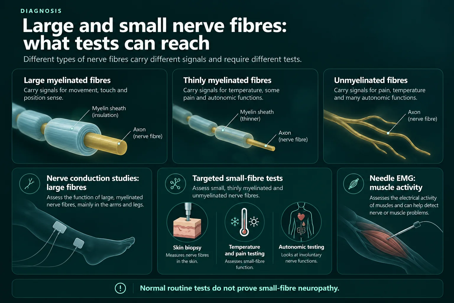
Large nerve fibres are thick and wrapped in myelin, an insulating layer that makes signals travel faster. Small fibres have only a thin layer of myelin or none at all. The autonomic fibres, which help run things you never think about, such as sweating, also count as small fibres. The table puts the two groups side by side; position sense means knowing where your body is without looking.1, 4, 5, 6, 12
| Your question | Large nerve fibres | Small nerve fibres |
|---|---|---|
| What are they? | Thick fibres with a myelin coat: motor fibres, and large sensory fibres (Aα and Aβ) | Thin fibres: thinly coated Aδ fibres and uncoated C fibres, plus the autonomic fibres |
| What do they do? | Motor fibres move your muscles. Aα and Aβ sensory fibres carry touch, vibration and position sense | Aδ and C fibres carry pain and temperature (Aδ: fast, well-localised pain; C: slower, less well-localised pain, and itch). Autonomic fibres help control things like sweating and blood flow in the feet |
| What can it feel like when they are affected? | Weakness, reduced vibration or position sense, unsteady walking, weaker tendon reflexes | Pain (often burning), changed temperature sense, pain from light touch; sometimes dry eyes or mouth, bowel or bladder symptoms, or dizziness when standing up |
| Do NCS and EMG see them? | Yes; results can be abnormal | No; results are normal if only the small fibres are affected |
| How else can they be checked? | Examination of strength, vibration sense and reflexes | Clinical examination for signs of small-fibre damage; in selected cases skin biopsy, quantitative sensory testing or autonomic tests |
| What does a normal NCS/EMG tell you? | No abnormality was found in the large fibres that were tested | Nothing either way; small fibre neuropathy needs positive findings of its own |
Why small fibres need other kinds of tests
Because NCS and EMG do not reach the small fibres, checking them relies on the clinical examination and, in some cases, specialised tests:5
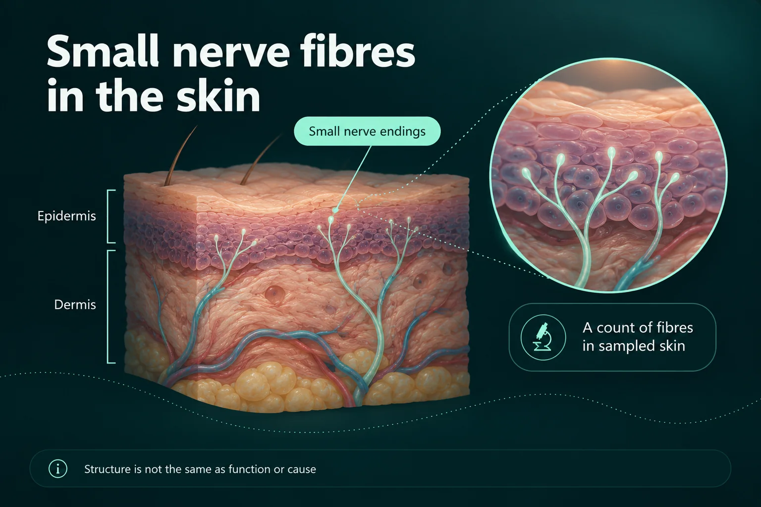
- Skin biopsy. A small skin sample, usually from the lower leg, in which the nerve fibres in the outer layer are counted and compared with expected values. A European guideline from 2010 calls it reliable when agreed counting rules and age-matched normal values are used. But reported sensitivity varies widely, so it does not catch every case, and it does not show what caused the damage.1, 7, 8
- Quantitative sensory testing (QST). Measures, among other things, when you feel warmth and cold. It depends on your own responses, and an international expert consensus on neuropathic pain (NeuPSIG) does not recommend it as a stand-alone test for diagnosis. In the Italian study, it was less accurate than skin biopsy.3, 9
- Autonomic tests, such as tests of sweating.5, 7
I want to be clear about one thing, because it is easy to hope otherwise. The sources describe skin biopsy as a test for people in whom small fibre neuropathy is suspected, not as a routine test for everyone with symptoms and a normal nerve conduction study. Whether more testing makes sense depends on what your doctor finds on examination and on the overall picture, not on symptoms alone. A 2023 Canadian review also notes that access to it varies widely in Canada.1, 3, 4
NCS and EMG are still useful when small fibre neuropathy is suspected: they can show whether large fibres are involved too, and point towards other explanations, such as a nerve root problem in the lower back.6
A normal result has limits, but it is not evidence of small fibre neuropathy
This is the part I think matters most. The limits of NCS and EMG explain why a small-fibre problem can be missed, but they do not turn a normal result into a positive finding. A normal test plus ongoing symptoms can look like proof of small fibre neuropathy. That is not how the diagnostic criteria work. In published criteria, a normal nerve conduction study is one of the conditions for calling a neuropathy “pure” small fibre neuropathy, but it is required in addition to positive findings, not instead of them. Those criteria ask for at least two abnormal findings among clinical signs of small-fibre damage, abnormal temperature thresholds on QST, and a reduced nerve fibre count on skin biopsy.3, 6
Symptoms on their own are not a reliable guide either. In the same Italian study, most people who had sensory symptoms but no clinical signs had normal small-fibre tests. And for the common type of polyneuropathy that starts in the feet, a case definition from the American Academy of Neurology and partner societies describes the combination of symptoms, examination findings and nerve test results as the most accurate basis for diagnosis.3, 10
I know that can be hard to read if you were hoping a test would finally give you an answer. But knowing what a result really says helps the next conversation with a doctor start from solid ground.
What else could be going on?
Symptoms with a normal test can also have other causes. Depending on the pattern, your doctor may think about:
- Nerve root problems in the lower back, such as lumbar radiculopathy, which can cause burning and numbness in the legs. Nerve root problems are typically uneven between the two sides, follow the skin area supplied by one root, and may come with neck or lower back pain.4, 6
- Conditions in the spinal cord or brain. An early question is whether symptoms come from the peripheral nerves or from the central nervous system. A disorder of the spinal cord in the neck (cervical myelopathy) is one condition that can mimic polyneuropathy.1, 4
How a test is done and read matters too. Results are compared with reference values that depend on factors such as age and height, and skin temperature affects the measurements: cooling slows nerve conduction, which is why your limbs are kept warm during testing.10, 11
Questions to take to your doctor about your own result
Which nerves and muscles are tested depends on what the test is meant to look for, so a “normal” report makes most sense in context.2 Questions you could bring to the doctor who requested or interpreted your test:
- Which nerves and muscles were tested, and what question was the test meant to answer?
- Were any results borderline, reduced or absent, rather than clearly normal?
- Do my examination findings point to large fibres, small fibres or neither?
- Could my symptoms come from somewhere other than the peripheral nerves, such as the spine or nerve roots?
- Is any further assessment of small fibres relevant in my case, and if not, why not?
- Would repeating the test make sense if my symptoms change?
I’m not a doctor. I’ve written this from published research and guidance, and to help you prepare questions for your own care. It can’t replace a doctor’s assessment.
Sources
- Mirian A, Aljohani Z, Grushka D, Florendo-Cumbermack A. Diagnosis and management of patients with polyneuropathy. CMAJ 2023;195(6):E227–E233, 13 February 2023. https://www.cmaj.ca/content/195/6/E227
- Horowitz SH; NeuropathyCommons (Mass General Brigham). Electromyography (EMG) and Nerve Conduction Studies. NeuropathyCommons, © 2022. https://neuropathycommons.org/diagnosis/electromyography-nerve-conduction
- Devigili G, Rinaldo S, Lombardi R, et al. Diagnostic criteria for small fibre neuropathy in clinical practice and research. Brain 2019;142(12):3728–3736. https://academic.oup.com/brain/article/142/12/3728/5610119
- Azhary H, Farooq MU, Bhanushali M, Majid A, Kassab MY. Peripheral Neuropathy: Differential Diagnosis and Management. American Family Physician 2010;81(7):887–892, 1 April 2010. https://www.aafp.org/pubs/afp/issues/2010/0401/p887.html
- Terkelsen AJ, Karlsson P, Lauria G, Freeman R, Finnerup NB, Jensen TS. The diagnostic challenge of small fibre neuropathy: clinical presentations, evaluations, and causes. Lancet Neurology 2017;16(11):934–944 (abstract). https://pure.au.dk/portal/en/publications/the-diagnostic-challenge-of-small-fibre-neuropathy-clinical-prese/
- Cascio MA, Mukhdomi T. Small Fiber Neuropathy. StatPearls Publishing (NCBI Bookshelf), updated 12 December 2022. https://www.ncbi.nlm.nih.gov/books/NBK582147/
- NeuropathyCommons (Mass General Brigham). Diagnosing Peripheral Neuropathy. NeuropathyCommons, © 2022. https://neuropathycommons.org/neuropathy/tests-diagnosing-small-fiber-polyneuropathy
- Lauria G, Hsieh ST, Johansson O, et al. European Federation of Neurological Societies/Peripheral Nerve Society guideline on the use of skin biopsy in the diagnosis of small fiber neuropathy. Journal of the Peripheral Nervous System 2010;15(2):79–92 (abstract). https://pure.johnshopkins.edu/en/publications/european-federation-of-neurological-societiesperipheral-nerve-soc-37
- Backonja MM, Attal N, Baron R, et al. Value of quantitative sensory testing in neurological and pain disorders: NeuPSIG consensus. Pain 2013;154(9):1807–1819 (abstract). https://experts.umn.edu/en/publications/value-of-quantitative-sensory-testing-in-neurological-and-pain-di/
- England JD, Gronseth GS, Franklin G, et al. Distal symmetrical polyneuropathy: a definition for clinical research. Muscle & Nerve 2005;31:113–123 (AAN/AANEM/AAPM&R report). https://www.aanem.org/docs/default-source/documents/distal-symmetrical-polyneuropathy-definition-for-clinical-research.pdf?sfvrsn=da35c6c2_1
- Ramani PK, Lui F, Arya K. Nerve Conduction Studies and Electromyography. StatPearls Publishing (NCBI Bookshelf), updated 10 February 2025. https://www.ncbi.nlm.nih.gov/books/NBK611987/
- Chen J, Kandle PF, Murray IV, Fitzgerald LA, Sehdev JS. Physiology, Pain. StatPearls Publishing (NCBI Bookshelf), updated 24 July 2023. https://www.ncbi.nlm.nih.gov/books/NBK539789/
Sources checked on 24 September 2026.
Related reading

Restless legs or neuropathy? Similar symptoms, different assessments
Written by someone living with neuropathy: how restless legs syndrome differs from neuropathy, why the two are so often confused, how often they occur together, how each is assessed, and questions to take to your doctor.

Cold feet: nerve symptoms, circulation and when to seek help
Written by someone living with neuropathy: why feet can feel cold without being cold, when cold feet point to circulation instead, the warning signs that need urgent help, and a description aid for your appointment.

Numb hands and weak grip: neuropathy or another nerve problem?
Written by someone living with neuropathy: why numb hands and a weak grip can be a trapped nerve, a neck problem or polyneuropathy, the warning signs, and a description aid to take to your doctor.