
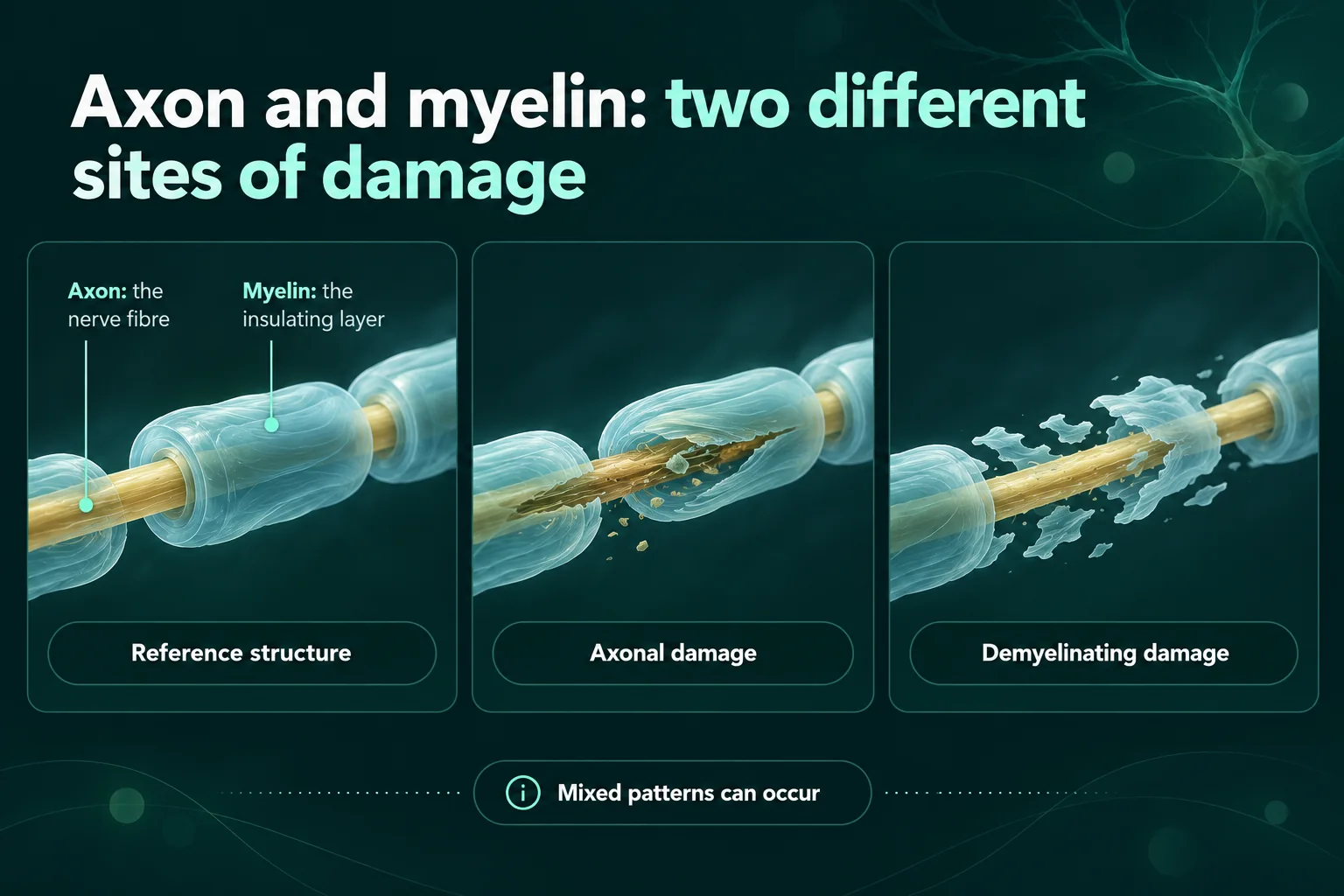
Axonal vs demyelinating neuropathy is about which part of the nerve fibre is damaged. Axonal means the nerve fibre itself, the axon, is damaged or lost. Demyelinating means the insulating layer around it, the myelin, is damaged. On a nerve conduction report, axonal damage mainly shows up as smaller signals, while demyelinating damage mainly shows up as slower signals. The pattern helps narrow down possible causes, but it does not decide the cause on its own.1, 2, 3
I have neuropathy myself, and test reports are full of words that are easy to misread when you are alone with them at the kitchen table. This page is a plain-language guide to those words, so that you can bring better questions to the person who ordered your test.
What do “axonal” and “demyelinating” mean?
A nerve fibre has two main parts. The axon is the long, thin extension of a nerve cell that carries the signal. Many axons are wrapped in myelin, an insulating layer. When the myelin is damaged, signals travel more slowly. The US National Institute of Neurological Disorders and Stroke (NINDS) says nerve tests can help show whether symptoms come from damage to the axon or to the myelin sheath.2, 3
I find it helpful to think of an electrical cable: the axon is the copper wire and the myelin is the plastic coating. If wires are cut or missing, less signal gets through. If the coating is damaged, the signal still gets through, but more slowly, or it may get stuck at the damaged spot. That picture is my own simplification, not a medical definition.
A review in the Canadian Medical Association Journal (CMAJ) explains that nerve conduction studies and electromyography (EMG) let the specialist sort a polyneuropathy as mainly axonal or demyelinating, as motor, sensory or both, and as acquired or inherited.1
Axonal vs demyelinating neuropathy on a nerve conduction report
In a nerve conduction study, small electrical pulses are given to a nerve and the response is recorded, for example over a muscle. The American Association of Neuromuscular & Electrodiagnostic Medicine (AANEM) describes it as showing how well the body’s electrical signals are travelling to a nerve. The table sets out the measurements you are most likely to see in a report.5, 6
| Word in the report | What it measures | Typical axonal pattern | Typical demyelinating pattern |
|---|---|---|---|
| Amplitude (SNAP for sensory nerves, CMAP for motor nerves) | The size of the response, a rough measure of how many fibres are conducting | Reduced | Can be normal or reduced, and may drop further along the nerve where the signal is blocked |
| Conduction velocity | How fast the signal travels, in metres per second | Normal or near normal | Slowed |
| Distal latency | The time from the pulse to the start of the muscle response, near the end of the nerve | Normal or near normal | Prolonged |
| F-wave latency | A response that travels up towards the spinal cord and back, so it also reflects the part of the motor nerve closest to the spine | Normal or only mildly prolonged | Prolonged |
| Conduction block | Whether many fibres fail to carry the signal past a short stretch of nerve | Not expected | Can be present, especially in acquired forms |
| Temporal dispersion | Whether signals arrive spread out in time because some fibres are slowed more than others | Not expected | Can be present, especially in acquired forms |
StatPearls, a medical reference on the US National Library of Medicine’s bookshelf, describes the classic axonal finding as reduced amplitudes with near-normal distal latency and conduction velocity. It describes the classic demyelinating findings as prolonged distal latency, slowed conduction velocity and prolonged F-wave latency, with conduction block and abnormal temporal dispersion as further signs.3
Two practical details are worth knowing. Cold limbs slow conduction, so the temperature of the arms and legs should be kept within a set range during the test. Normal values also vary, for example with age and height, and age-corrected values are used for young children and people over 60.6
Other words you may see in the report
- Sensory, motor or sensorimotor: whether the nerves for feeling, for movement, or both are affected.1
- Length-dependent: the longest nerves, such as those reaching the feet, are affected first. CMAJ calls this distal symmetric polyneuropathy and says it is the most common kind.1
- Mixed: features of both axonal and demyelinating damage. StatPearls describes the nerve test findings in diabetic neuropathy as typically a mixed axonal and demyelinating pattern.3
- Uniform or non-uniform slowing: in inherited demyelinating neuropathies the slowing is usually even and widespread, while conduction block and temporal dispersion point more towards an acquired cause. There are exceptions, such as one X-linked form of Charcot-Marie-Tooth disease.9
Why the picture is often mixed
The two kinds of damage do not stay neatly apart. StatPearls notes that as a mainly demyelinating neuropathy progresses, mild axonal damage may occur. In chronic inflammatory demyelinating polyradiculoneuropathy (CIDP), which is mainly demyelinating, axonal degeneration is commonly present as well, probably as a result of the inflammation.3, 10
So a report may say “predominantly axonal” or “mainly demyelinating” rather than one or the other. As I understand it, words like “predominantly” matter, and it is fair to ask what they mean for you.
What the pattern can point to
The pattern narrows down which causes are more or less likely. It is not a diagnosis. The Merck Manual lists diabetes, some chemotherapy drugs, alcohol and vitamin B12 deficiency among the causes of axonal neuropathy, and StatPearls describes the neuropathy after the chemotherapy drug cisplatin as mainly axonal. CMAJ describes chronic idiopathic axonal neuropathy, a slowly progressing neuropathy with no cause found, as typically starting after the age of 60.1, 3, 11
Demyelinating patterns point to a different, shorter list. It includes Guillain-Barré syndrome, CIDP and inherited neuropathies such as Charcot-Marie-Tooth disease type 1 (CMT1). The EAN/PNS guideline also lists anti-MAG neuropathy, which is linked to an abnormal antibody in the blood, among the conditions that can look like CIDP.3, 9, 12
In Charcot-Marie-Tooth disease, a median nerve conduction velocity of 38 metres per second is commonly used to separate the demyelinating from the axonal types.9
Does “demyelinating” mean I have CIDP?
No, not by itself. The joint European Academy of Neurology and Peripheral Nerve Society (EAN/PNS) guideline says the diagnosis of CIDP rests on a combination of clinical, electrodiagnostic and laboratory features, with other conditions that can look like CIDP ruled out. The guideline also says misdiagnosis commonly occurs, and that some people who fulfil the criteria based on correctly interpreted test results do not have CIDP.12
In a study by Allen and Lewis of 59 people referred with a diagnosis of CIDP, 47% did not meet the minimal diagnostic requirements. Liberal interpretation of what counts as demyelination on nerve tests was one of the common diagnostic errors they described.13
I find that reassuring rather than worrying: the word “demyelinating” is a reason to look carefully, not a label on its own.
Can the report be normal even if I have neuropathy?
Yes. Nerve conduction studies only test the larger nerve fibres. CMAJ says people with a neuropathy that affects only the small fibres will have normal findings, and StatPearls says small fibre problems are diagnosed with other tests, such as a skin biopsy. I cover this in more detail in my article on normal EMG and nerve conduction results.1, 14
Timing matters too. In Guillain-Barré syndrome, the results may be normal or non-specific when the test is done early in the illness, for example within the first week, and a repeat test may be needed.15
Does axonal or demyelinating tell me my outlook?
I have not found any source that predicts one person’s outlook from a single word in a report, and I would be careful with anyone who does. The cause, how long the damage has been going on and whether the cause can be treated all matter.
In general terms, a reference chapter on nerve injuries describes axons regrowing at roughly one millimetre a day, and the mildest injuries, which mainly affect the myelin, as recovering within days to weeks. Those figures describe injured nerves, not polyneuropathy, so they don’t translate directly to a report like yours. For polyneuropathy, the Merck Manual notes that even when a cause such as a medicine, a toxin or a vitamin deficiency is dealt with, recovery is slow and may be incomplete.11, 16
Who interprets the report, and what can you ask?
A nerve conduction report is not meant to be read in isolation. AANEM says the interpretation of the test results is an integral part of the examination, and that the conclusions come from combining the test results with the history and physical examination. Cleveland Clinic makes the same point more simply: nerve conduction studies alone don’t usually provide a diagnosis.17, 18
These questions may help at your next appointment:
- Is my neuropathy mainly axonal, mainly demyelinating, or mixed?
- Which nerves were affected: sensory, motor, or both?
- Is the pattern length-dependent, starting in the feet?
- Were there signs of conduction block or uneven slowing, and what do they suggest?
- Does the pattern change which causes you want to look for, or which further tests I need?
- Would a repeat test later be useful, and why?
I’m not a doctor. I’ve written this from published research and guidance, and to help you prepare questions for your own care.
Sources
- Mirian A, Aljohani Z, Grushka D, Florendo-Cumbermack A. Diagnosis and management of patients with polyneuropathy. CMAJ 2023;195(6):E227–E233. doi:10.1503/cmaj.220936. https://www.cmaj.ca/content/195/6/E227
- National Institute of Neurological Disorders and Stroke (NINDS). Peripheral Neuropathy. Last reviewed 13 March 2026. https://www.ninds.nih.gov/health-information/disorders/peripheral-neuropathy
- Novello BJ, Pobre T. Electrodiagnostic Evaluation of Peripheral Neuropathy. StatPearls [Internet]. StatPearls Publishing; last update 30 January 2023. NBK563169. https://www.ncbi.nlm.nih.gov/books/NBK563169/
- National Institute of Neurological Disorders and Stroke (NINDS). Guillain-Barré Syndrome. Last reviewed 13 March 2026. https://www.ninds.nih.gov/health-information/disorders/guillain-barre-syndrome
- American Association of Neuromuscular & Electrodiagnostic Medicine (AANEM). Types of Tests (patient information). No date shown. https://www.aanem.org/clinical-practice-resources/patient-information/types-of-tests
- Ramani PK, Lui F, Arya K. Nerve Conduction Studies and Electromyography. StatPearls [Internet]. StatPearls Publishing; last update 10 February 2025. NBK611987. https://www.ncbi.nlm.nih.gov/books/NBK611987/
- Haustein D, Reeves E, Swartz C. Basic Electrodiagnostics for the Referring Physician. PM&R KnowledgeNow, American Academy of Physical Medicine and Rehabilitation. Published 10 September 2015, updated 16 August 2024. https://now.aapmr.org/the-electrodiagnostic-consultation-and-report/
- Bromberg MB. An electrodiagnostic approach to the evaluation of peripheral neuropathies. Phys Med Rehabil Clin N Am 2013;24:153–168. doi:10.1016/j.pmr.2012.08.020. https://doi.org/10.1016/j.pmr.2012.08.020
- Nagappa M, Sharma S, Taly AB. Charcot-Marie-Tooth Disease. StatPearls [Internet]. StatPearls Publishing; last update 22 June 2024. NBK562163. https://www.ncbi.nlm.nih.gov/books/NBK562163/
- Gogia B, Rocha Cabrero F, Khan Suheb MZ, Lui F, Rai PK. Chronic Inflammatory Demyelinating Polyradiculoneuropathy. StatPearls [Internet]. StatPearls Publishing; last update 4 March 2024. NBK563249. https://www.ncbi.nlm.nih.gov/books/NBK563249/
- Feldman AM. Polyneuropathy. Merck Manual Professional Version. Last updated May 2026. https://www.merckmanuals.com/professional/neurologic-disorders/peripheral-nervous-system-and-motor-unit-disorders/polyneuropathy
- Van den Bergh PYK, van Doorn PA, Hadden RDM, et al. European Academy of Neurology/Peripheral Nerve Society guideline on diagnosis and treatment of chronic inflammatory demyelinating polyradiculoneuropathy: Report of a joint Task Force – Second revision. Eur J Neurol 2021;28(11):3556–3583. doi:10.1111/ene.14959. https://onlinelibrary.wiley.com/doi/10.1111/ene.14959
- Allen JA, Lewis RA. CIDP diagnostic pitfalls and perception of treatment benefit. Neurology 2015;85(6):498–504. doi:10.1212/WNL.0000000000001833. https://www.neurology.org/doi/10.1212/WNL.0000000000001833
- Cascio MA, Mukhdomi T. Small Fiber Neuropathy. StatPearls [Internet]. StatPearls Publishing; last update 12 December 2022. NBK582147. https://www.ncbi.nlm.nih.gov/books/NBK582147/
- Bhatti KS, Maheshwary A, Sun CE. Guillain-Barre Syndrome. StatPearls [Internet]. StatPearls Publishing; last update 31 January 2026. NBK532254. https://www.ncbi.nlm.nih.gov/books/NBK532254/
- Nadi M, Dabbas W, Das JM. Peripheral Nerve Injury. StatPearls [Internet]. StatPearls Publishing; last update 15 February 2026. NBK549848. https://www.ncbi.nlm.nih.gov/books/NBK549848/
- American Association of Neuromuscular & Electrodiagnostic Medicine (AANEM). Reporting the Results of Nerve Conduction Studies and Needle EMG. Approved October 2005; updated December 2024. https://www.aanem.org/docs/default-source/documents/aanem/practice/rptresultsemgncs-pdf.pdf
- Cleveland Clinic. Nerve Conduction Study: What It Is, Procedure & Results. Last reviewed 2 March 2023. https://my.clevelandclinic.org/health/treatments/24821-nerve-conduction-study
Sources checked on 25 September 2026.
Related reading

Restless legs or neuropathy? Similar symptoms, different assessments
Written by someone living with neuropathy: how restless legs syndrome differs from neuropathy, why the two are so often confused, how often they occur together, how each is assessed, and questions to take to your doctor.

Cold feet: nerve symptoms, circulation and when to seek help
Written by someone living with neuropathy: why feet can feel cold without being cold, when cold feet point to circulation instead, the warning signs that need urgent help, and a description aid for your appointment.

Numb hands and weak grip: neuropathy or another nerve problem?
Written by someone living with neuropathy: why numb hands and a weak grip can be a trapped nerve, a neck problem or polyneuropathy, the warning signs, and a description aid to take to your doctor.