
A skin biopsy for small fiber neuropathy takes a tiny round piece of skin, about 3 mm across, usually from the lower leg about 10 cm above the ankle. A specialised laboratory counts the small nerve fibres in the outer layer of the skin and compares the number with normal values for your age. It can show whether those fibres are reduced. It cannot tell you why, and a normal result does not always rule the condition out.1, 2
I have neuropathy myself, and when I first read about this test I wanted to know what it involves and what the number on the report really means. This page is about the biopsy itself: how the sample is taken, how it is measured, what it can and cannot show, and what to ask. Small fiber neuropathy as a whole, its symptoms, causes and treatment, has its own page on this site.
What is a skin biopsy for small fiber neuropathy?
Small fiber neuropathy affects the thinnest nerve fibres, the ones that carry pain and temperature and help control things like sweating. Standard nerve conduction studies and EMG only test the larger fibres, so a review in the Canadian Medical Association Journal (CMAJ) notes that people with purely small-fibre neuropathy will have normal results on those tests.4
That is where the skin biopsy comes in. The fine nerve endings in the outer layer of the skin, the epidermis, are the far ends of these small fibres. In the laboratory, the sample is cut into thin sections and stained so the nerve fibres show up. The lab then counts the fibres that cross from the deeper skin into the epidermis and divides by the length of skin examined. The result is called the intraepidermal nerve fibre density, often shortened to IENFD, and is given as fibres per millimetre.2, 5
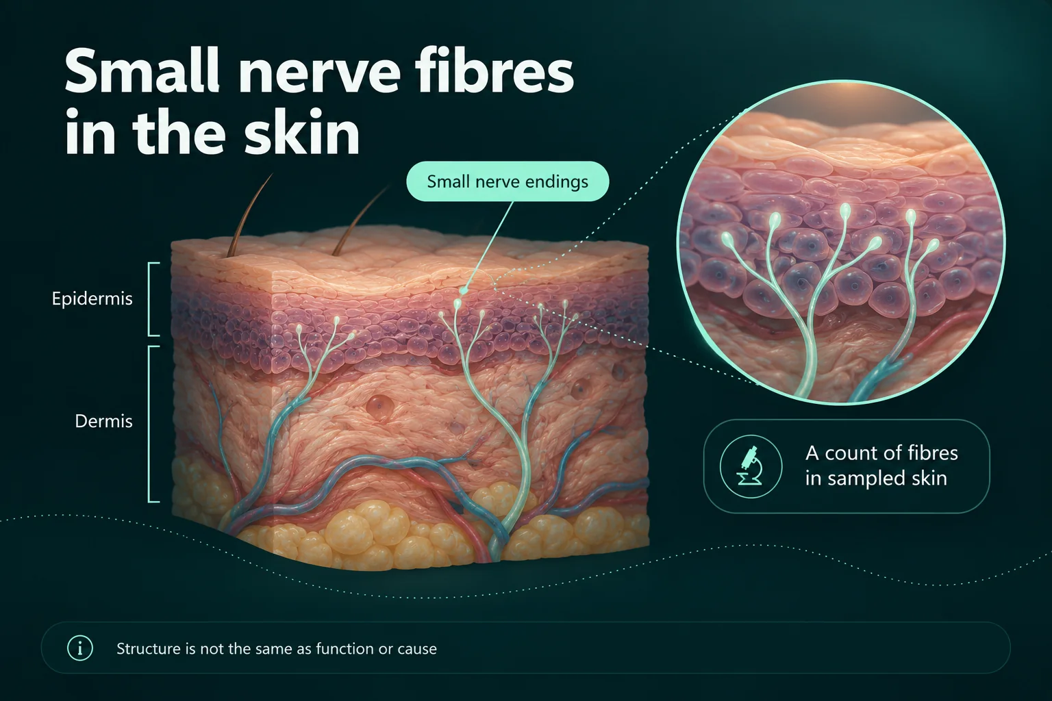
The European Federation of Neurological Societies and the Peripheral Nerve Society (EFNS/PNS) published a joint guideline in 2010. It gives a lower-leg biopsy with this count its strongest recommendation, level A, as a reliable and efficient way to assess the diagnosis. I could not find a newer version of that guideline, so as far as I can tell it is still the main reference.1
How is the sample taken?
The biopsy is a small outpatient procedure. Johns Hopkins’ cutaneous nerve laboratory says it takes 10 to 15 minutes and starts with a small injection of local anaesthetic to numb the skin. Cleveland Clinic’s protocol places the standard lower-leg sample on the outer side of the leg, 10 cm above the ankle bone, and the large international studies of normal values were collected at this lower-leg spot.6, 7, 8
Often more than one sample is taken. Mayo Clinic Laboratories normally asks for two samples from the same side of the body, and both Johns Hopkins and Cleveland Clinic describe three standard sites on one leg: the lower leg, the lower thigh and the upper thigh. The exact spots follow each lab’s own protocol. I come back to why a thigh sample can be useful further down.5, 6, 8
Cleveland Clinic’s protocol says no stitch is needed; the small wound is simply covered with a dressing. According to NeuropathyCommons, a patient resource from Mass General Brigham, the biopsy can be taken in almost any medical setting, often by dermatologists or neuromuscular specialists, and the sample is then sent to a special pathology laboratory.2, 8
Tell the person doing the biopsy if you take blood thinners. Johns Hopkins’ instructions for doctors say that people on anticoagulants with an INR above 2.5 should not have the biopsy. That is a decision for your care team; please don’t change or stop any medicine on your own because of a test.6
Does it hurt, and what are the risks?
The EFNS/PNS guideline calls the 3 mm ankle biopsy safe: ten laboratories reported no serious side effects in about 35,000 biopsies over roughly 15 years, and non-serious side effects in 0.19%.1
Like any skin biopsy, it can occasionally lead to bleeding, infection or a scar. MedlinePlus lists infection and scarring as risks of skin biopsies in general. Johns Hopkins describes the risk of bleeding or infection after this biopsy as extremely low.6, 9
If you have diabetes, poor circulation or slow-healing skin on your legs, I would mention it beforehand. I did not find a source that measures the healing risk for this biopsy in those groups, so this is my own suggestion rather than a documented finding.
Aftercare: looking after the small wound
Johns Hopkins’ patient information says to leave the dressings on and keep them dry for the rest of the day, that showering is fine from the next day with the plasters left on, and to avoid soaking in a bath or swimming while the wound heals. Johns Hopkins says healing usually takes one to two weeks. NeuropathyCommons suggests keeping the tight bandage on for a few hours, avoiding vigorous exercise for several hours, and keeping the area covered with a plaster until the scab drops off, which it says can take several weeks.2, 6, 10
NeuropathyCommons also says to call your doctor if redness or swelling gets worse or doesn’t go away, or if you see pus coming from the biopsy site.2
How is “normal” decided?
There is no single number that counts as normal for everyone. The EFNS/PNS guideline says the fibre density should always be compared with normal values matched for age. A worldwide study of 550 healthy people from eight laboratories found that the density falls with age in both women and men.1, 7
Sex and the exact biopsy spot matter too. In a multinational study of 528 healthy people from four laboratories, the lower limit of normal fell by 0.54 fibres per millimetre for each decade of age, was about 1.0 fibre per millimetre higher in women than in men, and was 3.4 fibres per millimetre lower for samples from the calf than from the standard spot on the outer lower leg.11
The thigh normally has more fibres than the ankle. In an early Johns Hopkins study of 98 healthy people, the average was about 21 fibres per millimetre at the thigh and about 14 at the lower leg. This is why each site has its own reference range.12
Staining methods differ as well, and the EFNS/PNS guideline asks new laboratories to set up their own age- and sex-matched normal values and quality checks. As I read it, a number on its own means little until you know the lab’s reference range for someone like you. A result is usually called reduced when it falls below the lab’s lower limit for your age and sex.1
When do the results come back?
Because the sample is processed in a specialised lab, the answer takes a while. Mayo Clinic Laboratories gives 7 to 14 days, Cleveland Clinic says a report generally reaches the referring doctor within two weeks of the lab receiving the samples, and Johns Hopkins says the turnaround is typically under three weeks.5, 8, 10
The report goes to the doctor who ordered the test. NeuropathyCommons notes that labs use different methods, and suggests asking which lab analysed your sample and getting a copy of the official report.2
What a skin biopsy can show
A reduced fibre density is objective evidence that the small nerve fibres in that patch of skin are fewer than expected. An Italian study screened 486 people referred with suspected sensory neuropathy and assessed 150 of them in detail. In that group, the skin biopsy had a diagnostic efficiency of 88.4%, compared with 54.6% for the clinical examination and 46.9% for quantitative sensory testing.13
Other studies give somewhat different figures, and the numbers are often quoted in different ways. A clinical review in the Cleveland Clinic Journal of Medicine gives a sensitivity of 88%, while a 2008 study of 99 people with neuropathy and 37 healthy controls, cited in a US insurer’s policy, found a sensitivity of 0.80 and a specificity of 0.82. I think “roughly 80 to 90%” is a fair summary, and it means the test is good but not perfect.14, 15
With a thigh sample as well, the biopsy can also show where along the leg the loss is greatest. A 2025 review explains that samples from the thigh allow doctors to assess the gradient of fibre loss. The same review notes that small fiber neuropathy usually starts furthest out, in the feet, and moves upwards, but that other patterns also occur.16
What decides the diagnosis is the whole picture. A 2019 study of 352 people with suspected sensory neuropathy, some of whom were followed over time, found that the skin biopsy was the most reliable tool to confirm small fiber neuropathy, but that combining clinical signs with a biopsy and/or sensory testing was more reliable than test results without clinical signs.17
What it cannot show
- The cause. The EFNS/PNS guideline says skin biopsy has so far not been useful for identifying the cause. To look for the cause, doctors may order blood tests and other investigations.1, 18
- How much it hurts. According to the same guideline, a reduced fibre density is linked to the risk of developing neuropathic pain but does not match its intensity.1
- How the fibres work. A 2025 review points out that the biopsy counts fibres but cannot assess how the small fibres function in the living body.16
- Every form of the condition. NeuropathyCommons explains that early, mild, recovering or patchy neuropathies may not severely damage the nerve endings in the exact spot biopsied, so some people with neuropathy still have a normal biopsy. A review on less typical forms describes small fiber neuropathy that does not start in the feet and can be patchy, uneven between the two sides, or mainly affect the arms.2, 19
According to Cleveland Clinic, about half of small fiber neuropathy cases happen for no known reason. A normal biopsy doesn’t mean your symptoms aren’t real, and a reduced one doesn’t tell you what comes next. Both are pieces of a larger conversation with your doctor.20
What about sweat gland nerves?
The same biopsy can also be used to look at the nerves around sweat glands. The EFNS/PNS guideline mentions a method for this, but a 2025 review says it is technically demanding and time-consuming and has not become routine. Aetna, a US insurer, considers it experimental.1, 15, 16
Other tests for small nerve fibres
A skin biopsy is not the only way to look at small fibres, and each test measures something different:
- Quantitative sensory testing (QST) measures when you first feel sensations such as warmth, cold or pain as a stimulus slowly changes, and you signal when you notice it. It depends on your full cooperation and understanding of the test.16
- Sweat tests such as QSART measure how much sweat the limbs produce in response to a substance called acetylcholine. The Cleveland Clinic Journal of Medicine describes QSART as a non-invasive autonomic test.14
- Other autonomic tests, such as heart rate and blood pressure responses, can support the diagnosis when relevant.18, 21
- Corneal confocal microscopy looks at the small nerves in the front of the eye. A 2024 review in ASRA Pain Medicine News says reduced corneal nerve density is associated with small fiber neuropathy.21
I’m not ranking these against each other. Which ones are available and useful depends on your symptoms and where you are treated. NeuropathyCommons puts it simply: no test is 100% accurate.18
Can I get a skin biopsy?
That depends on where you live and how your care is paid for. The CMAJ review calls skin biopsy the gold standard for small fiber neuropathy but says its availability and reimbursement vary widely across Canada. StatPearls, a reference for clinicians, notes that there is not yet a single gold standard test for the condition.4, 22
Insurers may also set conditions. Aetna’s policy, for example, considers the biopsy medically necessary for painful sensory neuropathy when nerve conduction studies and EMG are normal and certain other criteria are met, but considers it experimental for following the disease or the response to treatment over time. Other insurers and health systems have their own rules, so ask before the test if cost matters to you.15
The skin biopsy at a glance
This is my own summary of what the sources describe.
| Question | Short answer |
|---|---|
| Purpose | To count the small nerve fibres in the outer skin layer when small fiber neuropathy is suspected |
| How it is done | Local anaesthetic, one to three 3 mm punch samples from the lower leg and often the thigh, about 10 to 15 minutes, usually no stitches |
| Risks | Low; occasional bleeding, infection or a small scar |
| Results | Fibres per millimetre, compared with the lab’s normal values for age and sex; often one to three weeks |
| What it can show | Whether small fibres are reduced, and where along the leg |
| What it cannot show | The cause, how much it hurts, how the fibres function; a normal result can miss patchy or early changes |
Questions you can ask
- Why do you think a skin biopsy would help in my case, and what would change depending on the result?
- Which sites will you take samples from, and will one be from the thigh?
- Should I do anything about my blood thinners or other medicines, or leave them as they are?
- Which laboratory analyses the sample, and what are its normal values for my age and sex?
- When and how will I get the result, and can I have a copy of the report?
- If the result is normal, what else could explain my symptoms, and what happens next?
- Is the test covered, or will I pay for it myself?
I’m not a doctor. I’ve written this from published research and guidance, and to help you prepare questions for your own care.
Sources
- Lauria G, Hsieh ST, Johansson O, et al. European Federation of Neurological Societies/Peripheral Nerve Society Guideline on the use of skin biopsy in the diagnosis of small fiber neuropathy. Report of a joint task force of the EFNS and the PNS. J Peripher Nerv Syst 2010;15(2):79–92; also Eur J Neurol 2010;17(7):903–912. Abstract via Johns Hopkins research portal. https://pure.johnshopkins.edu/en/publications/european-federation-of-neurological-societiesperipheral-nerve-soc-37
- NeuropathyCommons, Mass General Brigham. Skin Biopsy. Undated (page copyright 2022). https://neuropathycommons.org/diagnosis/skin-biopsy
- National Institute of Neurological Disorders and Stroke (NINDS). Guillain-Barré Syndrome. Last reviewed 13 March 2026. https://www.ninds.nih.gov/health-information/disorders/guillain-barre-syndrome
- Mirian A, Aljohani Z, Grushka D, Florendo-Cumbermack A. Diagnosis and management of patients with polyneuropathy. CMAJ 2023;195(6):E227–E233. https://www.cmaj.ca/content/195/6/E227
- Mayo Clinic Laboratories. SPBX: Epidermal Nerve Fiber Density Consultation. Test catalog, undated. https://www.mayocliniclabs.com/test-catalog/overview/70599
- Johns Hopkins Medicine, Cutaneous Nerve Laboratory. For Physicians: Cutaneous Nerve Biopsy and Analysis. Undated. https://www.hopkinsmedicine.org/neurology-neurosurgery/specialty-areas/cutaneous-nerve-lab/physicians
- Lauria G, Bakkers M, Schmitz C, et al. Intraepidermal nerve fiber density at the distal leg: a worldwide normative reference study. J Peripher Nerv Syst 2010;15(3):202–207. doi:10.1111/j.1529-8027.2010.00271.x. Abstract via Maastricht University. https://cris.maastrichtuniversity.nl/en/publications/intraepidermal-nerve-fiber-density-at-the-distal-leg-a-worldwide-/
- Cleveland Clinic Neuromuscular Center. Skin Biopsy in the Diagnosis of Small Fiber Neuropathy – A Guide for Providers. Brochure 14-NEU-1564, 2014. https://my.clevelandclinic.org/-/scassets/files/org/neurological/neuromuscular/14-neu-1564-skin-biopsy-brochure-5.ashx?la=en
- MedlinePlus Medical Encyclopedia. Skin lesion biopsy. US National Library of Medicine; updated 2 August 2025. https://medlineplus.gov/ency/article/003840.htm
- Johns Hopkins Medicine, Cutaneous Nerve Laboratory. Cutaneous Nerve Biopsy Patient Information. Undated. https://www.hopkinsmedicine.org/neurology-neurosurgery/specialty-areas/cutaneous-nerve-lab/patients
- Provitera V, Gibbons CH, Wendelschafer-Crabb G, et al. A multi-center, multinational age- and gender-adjusted normative dataset for immunofluorescent intraepidermal nerve fiber density at the distal leg. Eur J Neurol 2016;23(2):333–338. doi:10.1111/ene.12842. Abstract via University of Minnesota. https://experts.umn.edu/en/publications/a-multi-center-multinational-age-and-gender-adjusted-normative-da-2
- McArthur JC, Stocks EA, Hauer P, Cornblath DR, Griffin JW. Epidermal nerve fiber density: normative reference range and diagnostic efficiency. Arch Neurol 1998;55(12):1513–1520. doi:10.1001/archneur.55.12.1513. Abstract via Johns Hopkins research portal. https://pure.johnshopkins.edu/en/publications/epidermal-nerve-fiber-density-normative-reference-range-and-diagn-3/
- Devigili G, Tugnoli V, Penza P, et al. The diagnostic criteria for small fibre neuropathy: from symptoms to neuropathology. Brain 2008;131(7):1912–1925. doi:10.1093/brain/awn093. https://academic.oup.com/brain/article/131/7/1912/384848
- Tavee JO. Office approach to small fiber neuropathy. Cleve Clin J Med 2018;85(10):801–812. https://www.ccjm.org/content/85/10/801
- Aetna. Nerve Fiber Density Measurement. Clinical Policy Bulletin 0774; last reviewed 11 December 2025. https://www.aetna.com/cpb/medical/data/700_799/0774.html
- Furia A, Liguori R, Donadio V. Small-Fiber Neuropathy: An Etiology-Oriented Review. Brain Sci 2025;15(2):158. doi:10.3390/brainsci15020158. https://www.mdpi.com/2076-3425/15/2/158
- Devigili G, Rinaldo S, Lombardi R, et al. Diagnostic criteria for small fibre neuropathy in clinical practice and research. Brain 2019;142(12):3728–3736. doi:10.1093/brain/awz333. https://academic.oup.com/brain/article/142/12/3728/5610119
- NeuropathyCommons, Mass General Brigham. Diagnosing Peripheral Neuropathy. Undated (page copyright 2022). https://neuropathycommons.org/neuropathy/tests-diagnosing-small-fiber-polyneuropathy
- Gemignani F, Bellanova MF, Saccani E, Pavesi G. Non-length-dependent small fiber neuropathy: not a matter of stockings and gloves. Muscle Nerve 2022;65(1):10–28 (published online 9 August 2021). doi:10.1002/mus.27379. Summary via AANEM education catalogue. https://education.aanem.org/Public/Catalog/Details.aspx?id=wMtT7sb55AiWKzIaDbSdlg%3D%3D
- Cleveland Clinic. Small Fiber Neuropathy: What It Is, Symptoms & Treatment. Last reviewed 13 November 2023. https://my.clevelandclinic.org/health/diseases/17479-small-fiber-neuropathy
- Levinson J, Collis R, Singla P. A Review of Small Fiber Neuropathy. ASRA Pain Medicine News, 6 November 2024. https://asra.com/news-publications/asra-newsletter/newsletter-item/asra-news/2024/11/06/a-review-of-small-fiber-neuropathy
- Cascio MA, Mukhdomi T. Small Fiber Neuropathy. StatPearls [Internet]. StatPearls Publishing; last update 12 December 2022. NBK582147. https://www.ncbi.nlm.nih.gov/books/NBK582147/
Sources checked on 26 September 2026.
Related reading

Restless legs or neuropathy? Similar symptoms, different assessments
Written by someone living with neuropathy: how restless legs syndrome differs from neuropathy, why the two are so often confused, how often they occur together, how each is assessed, and questions to take to your doctor.

Cold feet: nerve symptoms, circulation and when to seek help
Written by someone living with neuropathy: why feet can feel cold without being cold, when cold feet point to circulation instead, the warning signs that need urgent help, and a description aid for your appointment.

Numb hands and weak grip: neuropathy or another nerve problem?
Written by someone living with neuropathy: why numb hands and a weak grip can be a trapped nerve, a neck problem or polyneuropathy, the warning signs, and a description aid to take to your doctor.